Osteolysis
Home Up Materials Radiography Arthrography CT and MRI References


Home
Materials
Fixation
Prosthesis Types
 Total Hip 
 Hemi
 Revision 
Radiography
Baseline Exam
 Alignment
 -Acetab. Position
 -Inclination
 -Anteversion
 -Center of Rotation
 -Femoral Stem 
 -Leg Length Discr.
 Intra-op- Fractures 
 Cement Herniation
Follow-up Study
 Bony Abnormalities
 Fracture
 Heterotopic Bone
 Non Union
 Hardware Failure
 -Polyethylene Wear
 -Dislocation
 Osteolysis
 Loosening
 Infection
Arthrography
CT and MRI
References

Osteolysis

  • AKA particle disease, aggressive granulomatosis
  • Particulate debris from wear of arthroplasty components, most commonly polyethylene component (Schmalzried)
  • Large pieces sequestered in fibrous tissue
  • Small pieces taken up by macrophages and multinucleated giant cells which may release cytokines that initiate a cascade reaction ultimately resulting in osteolysis (Amstutz and Campbell)
  • Radiographs demonstrate focal areas of lytic bone
  • Large amounts of particulate may dissolve in joint fluid, staining the fluid and synovium a blackish color. This is termed metallosis.
  • Large areas can occur, do not necessarily indicate prosthesis loosening
  • Potential risk of pathologic fracture
  • May require revision surgery even in absence of symptoms due to danger of pathologic fracture and loss of bone increasing the difficulty of surgery


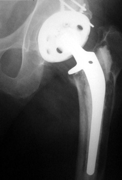
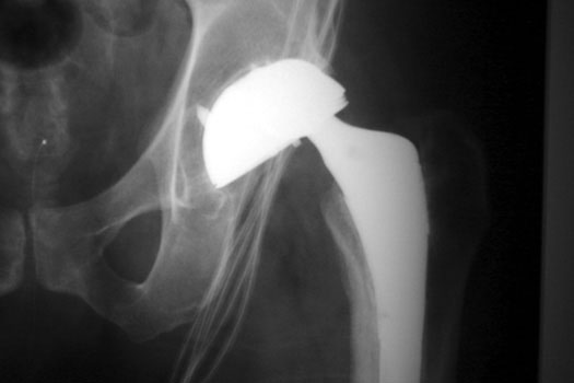
OSTEOLYSIS—Gruen zones 2 and 3, with markedly thinned femoral cortex, placing patient at risk for pathologic fracture


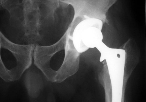
OSTEOLYSIS—ABOUT ACETABULAR COMPONENT GRUEN ZONE II, WITH PATHOLOGIC FRACTURE
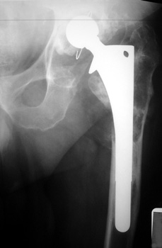
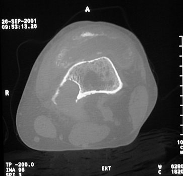
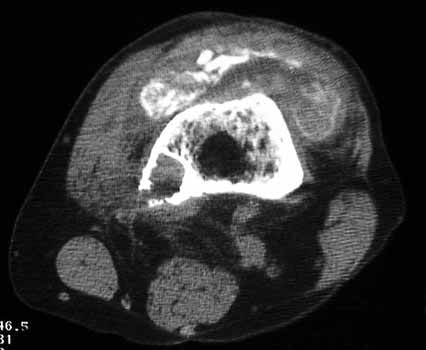

OSTEOLYSIS
Multifocal and massive, involving Gruen zone III of acetabular cup and Grune zone 1,2, and 6 of femoral component


OSTEOLYSIS
Radiograph and CT


OSTEOLYSIS
CT guided biopsy


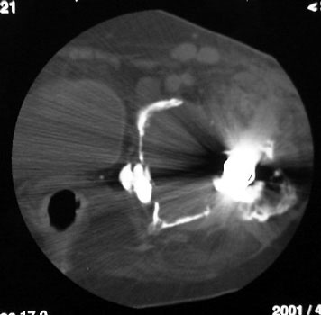
Osteolysis and metallosis in a patient with a total knee replacement. A focal area of osteolysis is seen in the posterior medial femur. Particulate debris is present throughout the suprapatellar bursa.


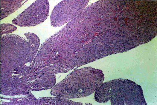
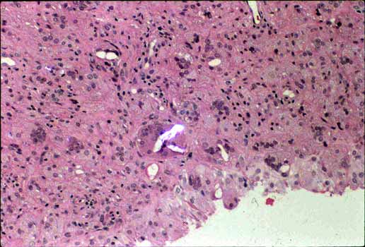
Prosthetic synovitis, low power. Hyperplastic synovial membrane obtained adjacent to prosthesis.

OSTEOLYSIS high power. Methylmethacralate debris (linear webs) surrounded by giant cells.

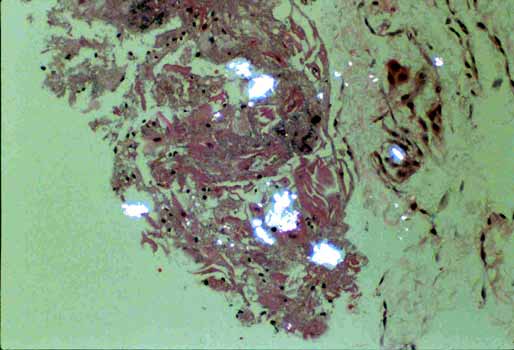
OSTEOLYSIS high power polarized microscopy. Polyethylene flakes (shiny linear material) surrounded by foreign body giant cell reaction. 

OSTEOLYSIS low power. Polyethylene flakes (shiny globular material) surrounded by foreign body giant cell reaction and histiocytes.


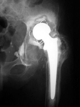
Dislocation simulating severe polyethylene wear or. The femoral head is posteriorly dislocated a just slightly projects superior to the cup


Google
 
Web Gentili.net