![]()
![]()
![]()
![]()
![]()
![]()
|
|
|
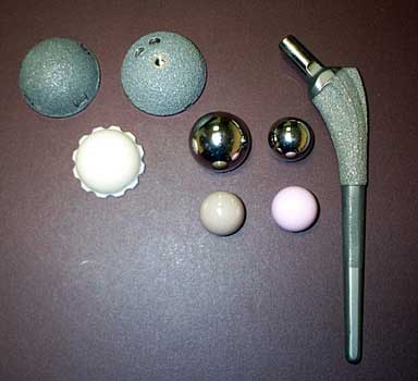
HIP ARTHROPLASTY:NORMAL AND ABNORMAL IMAGING APPEARANCESLakeside Center - Education Exhibits - Space 0101CE-e Thomas Learch MD, Amilcare Gentili MD, Deborah Forrester MD, Edward McPherson MD
Newer joint replacement systems and surgical techniques combined with the aging US population have lead to an increase in patients undergoing hip arthroplasty surgery. Baseline and follow-up imaging studies help in determining success or failure of joint replacement surgery, and aid in identifying common complications. Radiologists following these patients need to be aware of the normal radiographic appearance of post operative and follow up studies, as well as complications that occur over the course of implant use. This computer exhibit will describe the normal and abnormal radiographic appearances postoperatively and on follow up studies, including alignment and positioning abnormalities of the acetabular cup and femoral prostheses, bone, cement, and hardware fracture, polyethylene cup wear and dislocation, particle disease, loosening, and infection. Conventional radiography, arthrography, nuclear medicine, and multislice computed tomography imaging studies will be demonstrated. Imaging of antibiotic impregnated cement spacers used to treat infection as well as revision hip arthroplasty will also be presented.
|
|
|