Intra-op Fractures
Home Up Materials Radiography Arthrography CT and MRI References


Home
Materials
Fixation
Prosthesis Types
 Total Hip 
 Hemi
 Revision 
Radiography
Baseline Exam
 Alignment
 -Acetab. Position
 -Inclination
 -Anteversion
 -Center of Rotation
 -Femoral Stem 
 -Leg Length Discr.
 Intra-op- Fractures 
 Cement Herniation
Follow-up Study
 Bony Abnormalities
 Fracture
 Heterotopic Bone
 Non Union
 Hardware Failure
 -Polyethylene Wear
 -Dislocation
 Osteolysis
 Loosening
 Infection
Arthrography
CT and MRI
References

Intraoperative Fractures

  • Incidence
    • Cemented prostheses 0.4%
    • Press-fit prostheses 2.5%
    • Revision hip arthroplasty 7.2%
  • Location
    • Pelvis 
      • rare
    • Femoral shaft 
      • Most common
      • Usually at tip of stem in vertical configuration
      • Rarely displaced
      • Occur as the long femoral component stem is pounded into position
      • Usually does not effect outcome.
      • May require cerclage cables
  • DDx:
    • Nutrient foramen in posterior femoral shaft. Always compare with preoperative radiographs
    • Control perforation placed by surgeon during revision surgery to aid in removal of previously placed femoral component
  • Tx
    • Usually managed with cautious weight bearing

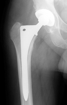
Intraoperative fracture of the proximal femur.


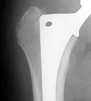
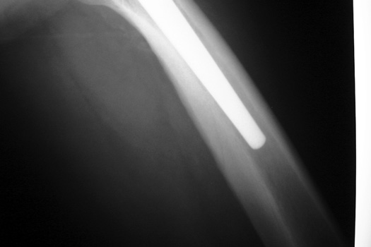
Intraoperative fracture of the femur at the tip of the femoral stem.


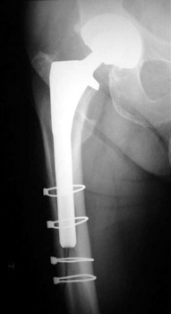
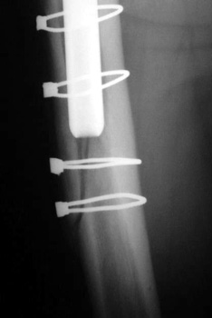
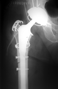
Femoral shaft fracture fixed with cerclage cables.


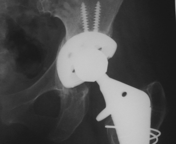
MEDIAL ACETABULAR FRACTURE


Control perforation lateral femoral cortex, reduced by cerclage cables. A control perforation of the lateral femoral cortex was performed in this revision total hip replacement to facilitate removal of the old femoral prosthesis. It is reduced by cerclage cables.


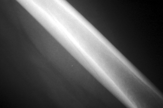
Vascular channel. Linear lucency seen on pre and post operative frog views is vascular channel. These are seen best on the frog leg view, entering the femoral cortex distally and traveling proximally toward the hip into the marrow space.


Google
 
Web Gentili.net