![]()
![]()
![]()
![]()
![]()
![]()
|
|
|
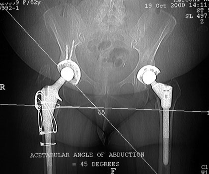
CT and MRIPreviously, CT and MRI were rarely used for patients with arthroplasties due to metallic artifact. Multislice CT has markedly decreased this artifact, and can visualize bone immediately adjacent to hardware. It can be used to evaluate alignment abnormalities, bone graft, heterotopic ossification, osteolysis, and adjacent soft tissue structures. Reformatted images in various planes can be generated with minimal artifact as well. MRI can also be used in patients with hardware by applying sequences that minimize metallic artifact. These sequences include T1 and STIR, employing long echo train lengths. Gradient echo and fat saturation techniques are avoided when hardware is present. ACETABULAR COMPONENT Acetabular component lateral inclination
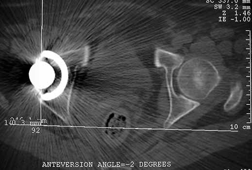
ACETABULAR COMPONENT Cup Anteversion
Cup in neutral position
Anteverted cup
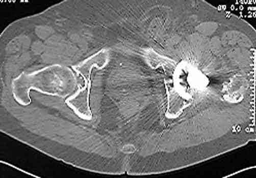
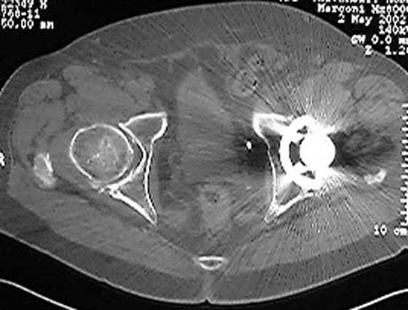
Femoral version measure by CT. Axial images are obtained through the hips and femoral condyles. A reference line is drawn through the posterior aspect of the medial and femoral condyles (Fig A). A second line in drawn through the axis of the neck of the femoral prosthesis. These lines are superimposed, and the femoral version angle is measured. (Fig B)
Hernia
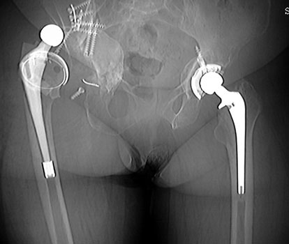
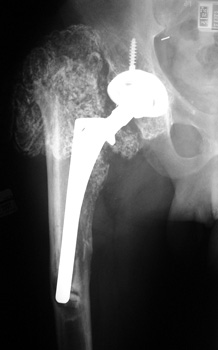
Dislocated femoral component and acetabular cup in grossly loose arthroplasty. CT guided aspiration to rule out infection. OSTEOLYSIS
HETEROTOPIC BONE FORMATION plain film and CT
CT—Bone graft placed for patient with congenital hip dysplasia and poorly developed acetabulum (Shelf procedure). Persistent cleft between graft and native bone is present years after surgery, consistent failure of graft incorporation
C
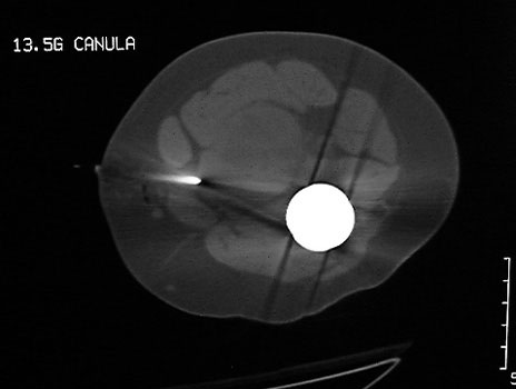
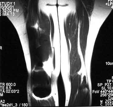
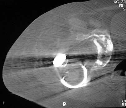
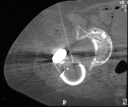
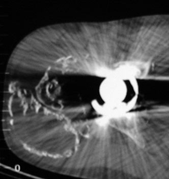
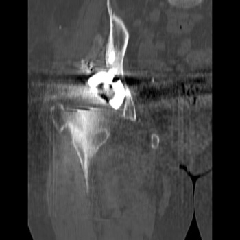
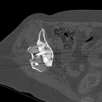
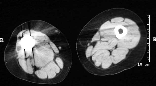
Mass in patient with modular endoprosthesis placed after
resection of femur for osteosarcoma. Axial (fig A) and coronal reformatted
images (fig B) clearly demonstrate mass adjacent to modular endoprosthesis. 14 gauge
core needle biopsy performed under CT guidance (fig C) demonstrated
recurrent osteosarcoma
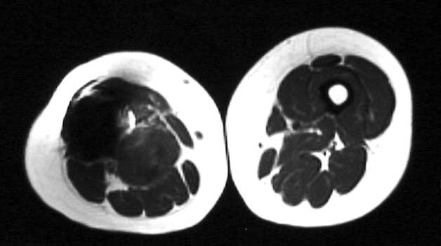
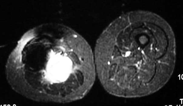
Recurrent osteosarcoma in patient status post resection of femur and placement of modular endoprosthesis. Axial T1 and STIR sequences clearly demonstrate mass adjacent to endoprosthesis. |
|
|
 B
B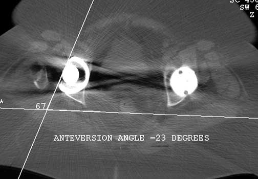
 A
A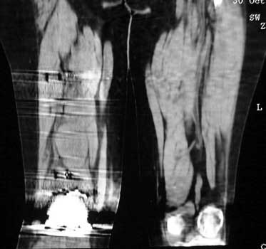 B
B