The word gangrene comes from the Latin word gangraena, an eating sore. Gangrene
is death and decay of a body part due to deficiency of blood supply and is frequent
complication in the diabetic foot.
Dry gangrene can be diagnosed on conventional radiographs, especially in the toes, when
marked thinning of the soft tissue shadows is present. MR imaging is more dramatic with
marked signal loss on all sequences secondary to absence of mobile protons necessary for
signal. Administration of intravenous gadolinium may show abrupt signal loss at the level
of the gangrenous change. Bone marrow signal changes may be present suggesting
osteomyelitis. This finding may be secondary to coexistent osteomyelitis or to ischemic
changes in the bone. Differentiation of these two entities cannot be accomplished by MR
imaging.


Gangrene
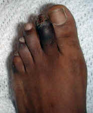
Two patients with dry gangrene of toes.
21-year-old man with newly diagnosed diabetes. Photograph of foot demonstrates
dry gangrene of the second toe.
50-year-old man with diabetes. PA radiograph demonstrates marked soft tissue
thinning of the 2nd and 3rd digits. The osseous structures, however, are intact without
evidence of destructive change.
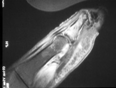
Sagittal MR images of 1st toe with wet gangrene. T1 and STIR images demonstrate lack of
signal distally. Sagittal T1 weighted images with fat saturation post intravenous
gadolinium demonstrate extensive tissue necrosis.
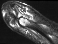
 52-year-old man with
long standing
diabetes and black 1st toe. Sagittal T1 weighted image with fat saturation
post intravenous gadolinium enhancement demonstrates absent signal of the distal soft tissues and bone of the 1st toe
secondary to dry gangrene. On
comparison radiographs, the 1st distal phalange was intact and present. On
MR images, the distal phalange is difficult to image due to lack of mobile
protons in dry gangrenous tissues. (Click on the images to see larger
versions)
 A
A  B
B
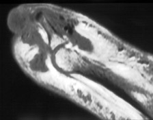
61-year-old man with diabetes and wet gangrene. Sagital STIR (A) and sagital T1
weighted image with fat saturation post intravenous gadolinium enhancement (B) demonstrate
extensive soft tissue necrosis. Lack of gadolinium enhancement of this area is consistent
with soft tissue necrosis and gangrene. (Click on the images to see larger
versions)