![]()
![]()
![]()
![]()
![]()
![]()
Atlas of Signs in Musculoskeletal Radiology is approved by the ARRS (American Roentgen Ray Society) and is included in AJR Webreview
A. Gentili,MD, M. Beller, MD, S. Masih, MD, L.L. Seeger, MD
![]()
|
Atlas of Signs in Musculoskeletal Radiology is approved by the ARRS (American Roentgen Ray Society) and is included in AJR WebreviewA. Gentili,MD, M. Beller, MD, S. Masih, MD, L.L. Seeger, MD
|
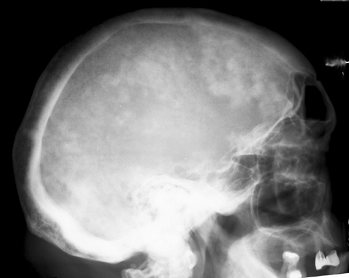
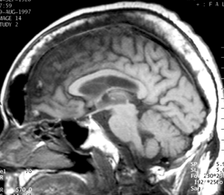
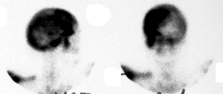
Diagnosis:Paget's Disease Discussion:Plain film of the skull reveals a large mottled area of radiolucency with small areas of increased density within it. The MR image of the skull reveals a thickened, enlarged cranium with increase in the marrow space. Two bone scan images also reveal increased activity in the skull, more localized to one side, characteristic to the localized disease seen in Paget's. This is classic cranial involvement of Paget's. In the cranium, bone sclerosis may produce circular radiodense lesions in one area, whereas osteoporosis circumscripta is noted elsewhere. In the skull, the common region of involvement is the cranial vault. The osteolytic phase is called osteoporosis circumscripta and appears as multiple geographic, well-demarcated regions of bone resorption that may be mistaken for metastases. Focal radiodensities occur as pagetoid bone is formed. In the quiescent phase, there is a radiodense cotton-wool appearance with a thickened vault.
References:
|
|
Atlas of Signs in Musculoskeletal Radiology is approved by the ARRS (American Roentgen Ray Society) and is included in AJR WebreviewA. Gentili,MD, M. Beller, MD, S. Masih, MD, L.L. Seeger, MD
|