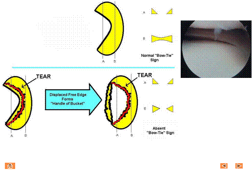
Bow-tie sign
(Adapted from Vahlensieck et al.
& Helms et al.)
Central
sagittal slice sequences of the normal meniscus demonstrate triangles of signal void (above,
A). On peripheral sagittal sequences,
the normal
meniscus has a “bow tie” appearance (above, B) for 2-3 consecutive slices. With a displaced tear, normal bow-tie
configuration is absent on consecutive sagittal sequences (below, B).
Lat Menisc
Tibia
Lat
Femoral Condyle
Normal bow-tie configuration of the lateral meniscus posterior horn is demonstrated (above).