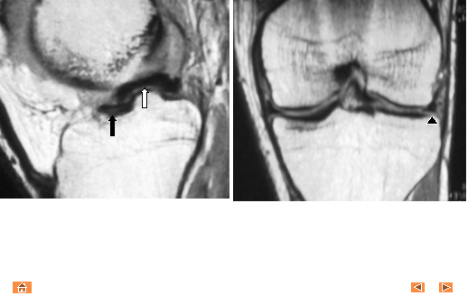

Coronal and sagittal T1 MR images demonstrate posterior
horn of the lateral?? meniscus (white arrow) flipped anterior, and superior to the
ipsilateral anterior horn (black arrow). Note
the diminutive residual lateral meniscus (black arrowhead).
Flipped Meniscus sign