
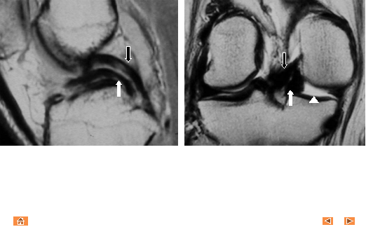
Double PCL sign
Sagittal and coronal proton-density MR images demonstrate
displaced medial meniscal fragment in the intercondylar notch (white arrow),
anterior to the posterior cruciate ligament (PCL, black arrow). Note that the displaced meniscal fragment lies in the same sagittal plane as the
PCL. Normal triangular shape of the peripheral medial meniscus is
truncated (white arrowhead).