|
1
|
|
|
2
|
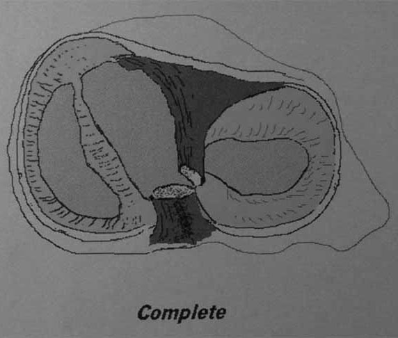
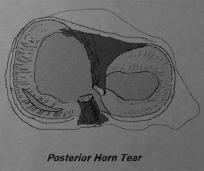
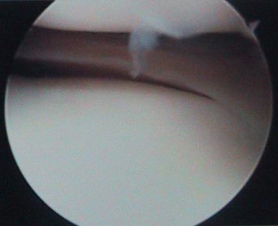
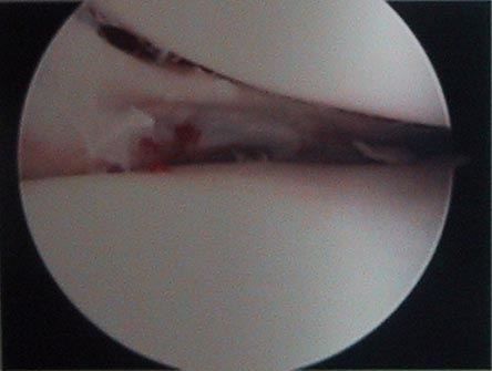
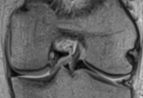
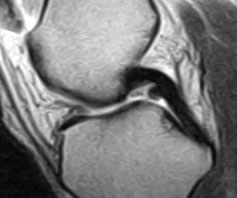
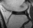
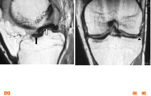
- Pathology: Bucket handle tear
represents a long-axis longitudinal tear of the meniscus (bucket), and
displacement of the attached inner fragment (handle).
- Commonly, these tears involve the medial meniscus, begin with a
vertical or oblique posterior horn tear, and propagate anteriorly and
longitudinally. 10% of meniscal
tears are of a bucket handle configuration.
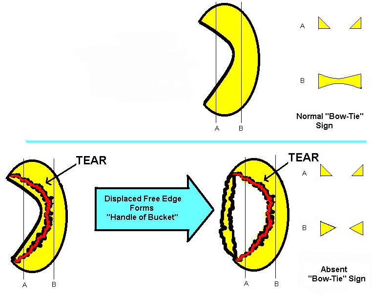
- Imaging findings: Frequently,
disruption of the normal Bow-tie sign configuration of the peripheral
meniscus is noted (sagittal view).
- Note: Bucket handle tears are commonly overlooked, because of the
parallel orientation of the tear on sagittal view, and similarity to the
joint space on coronal view.
Specific signs for associated variants of meniscal bucket handle
tears include:
- Flipped Meniscus sign
- Fragment-in-Notch sign
- Double PCL sign
- Double ACL sign
- Pitfalls: In addition, potential
pitfalls which may be confused with meniscal tears are discussed.
|
|
3
|
|
|
4
|
|
|
5
|
|
|
6
|
|
|
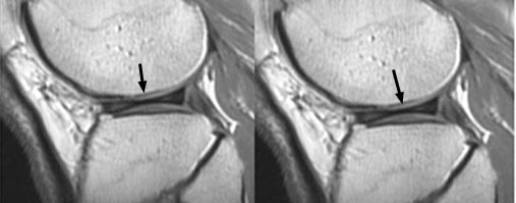
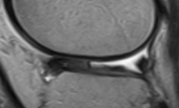
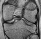
7
|
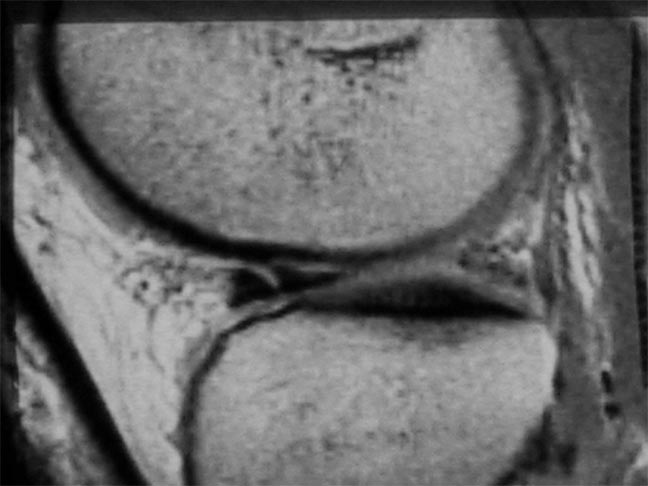
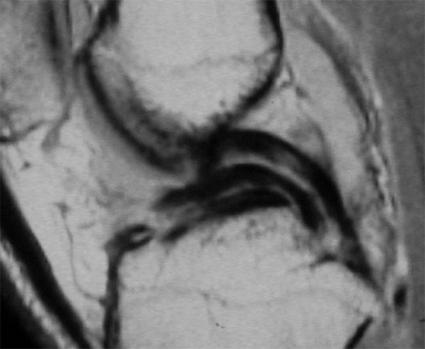
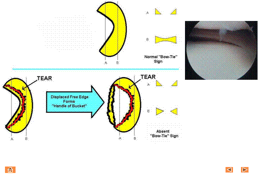
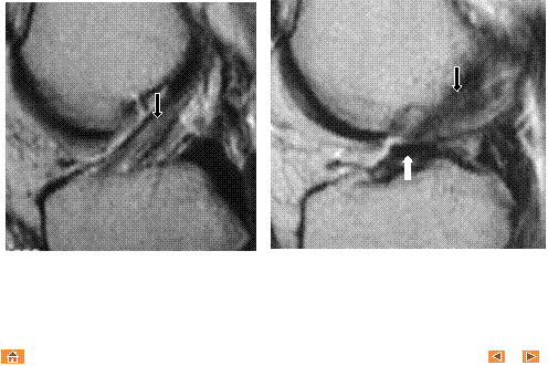
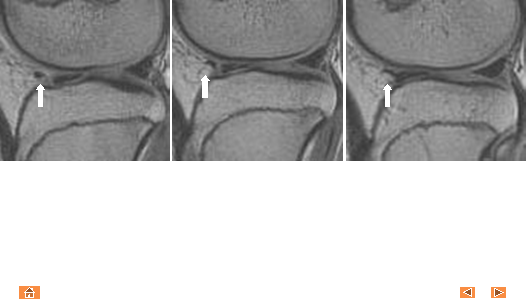
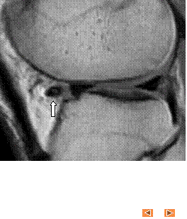
- Multiple sagittal T1 weighted images reveal loss of the normal bow-tie
appearance of the meniscus (black arrow). The "absent bow tie
sign" is another good sign of a bucket handle tear of the meniscus.
The absence of the normal bow-tie is secondary to the displaced fragment
which makes up the "handle" of the bucket. Requirement for the
absent bow tie sign mandates that the normal requirement of at least two
adjacent sagittal images with a normal meniscal body segment appearance
is not present.
|
|
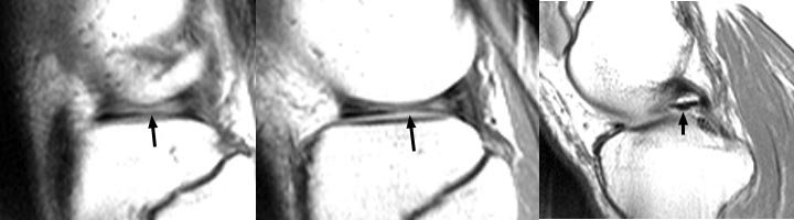
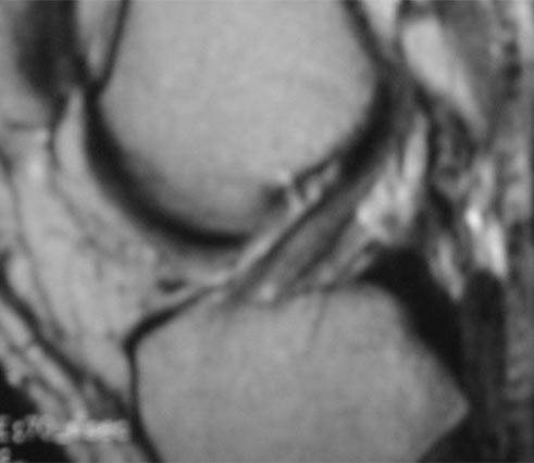
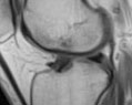
8
|
- Pathology: Inner fragment of torn
meniscus is flipped over ipsilateral anterior horn, and lies superior or
posterior to the normal anterior horn.
Occurs more frequently with medial, rather than lateral, meniscal
tears.
- Imaging findings: Abnormally
large anterior meniscus (8+ mm), measured in vertical dimension, with a
diminutive posterior horn (sagittal view). Alternatively, anterior lying fragment
results in elongated anterior horn with a band of high signal intensity
differentiating the anterior horn from the immediately adjacent
(anterior or posterior) flipped meniscal fragment.
- Note: The flipped meniscus is frequently associated with concurrent
fragment-in-notch meniscal displacement.
|
|
9
|
|
|
10
|
|
|
11
|
|
|
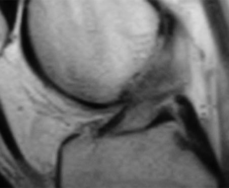
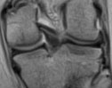
12
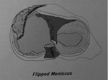
|
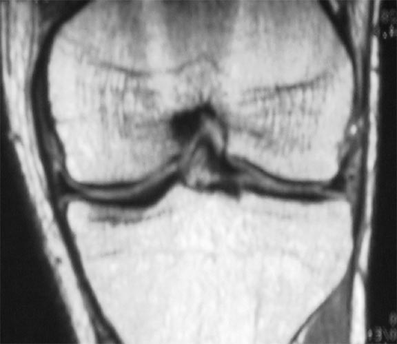
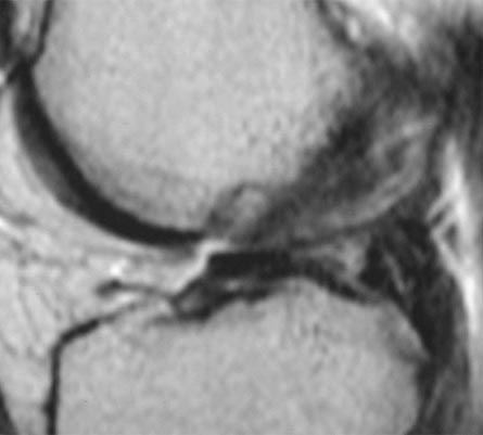
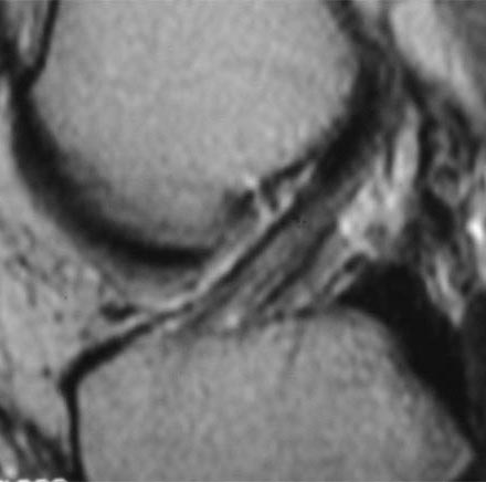
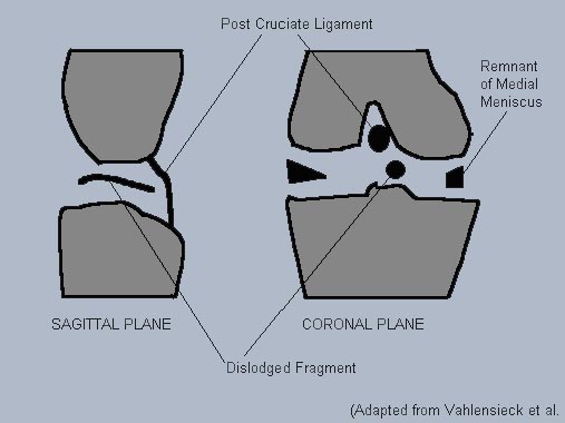
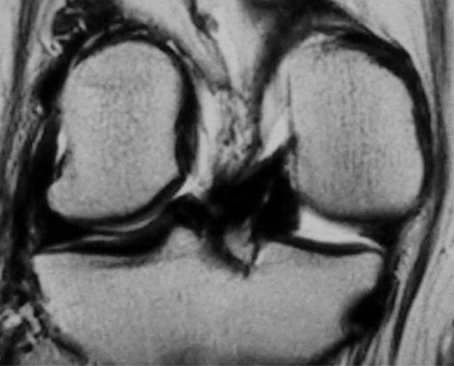
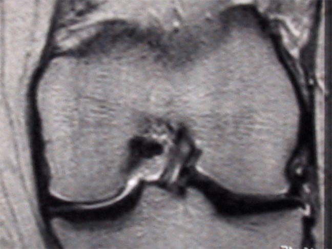
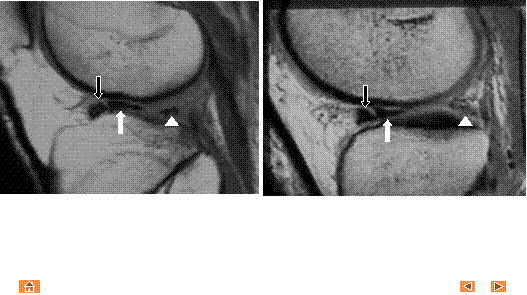
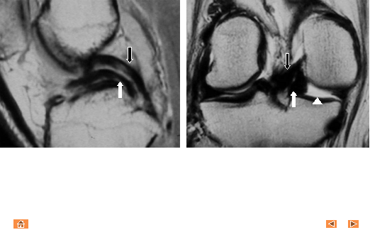
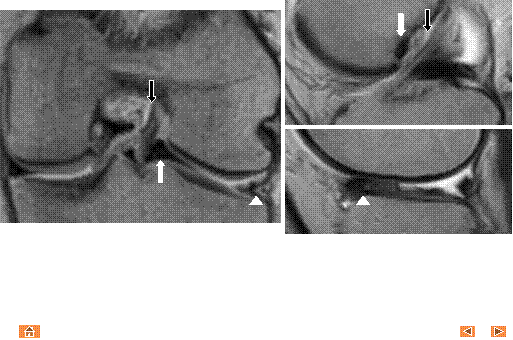
- Pathology: Meniscal bucket handle
tear, with meniscal fragment displaced within the intercondylar notch.
Occurs more frequently with medial, rather than lateral, meniscal tears.
- Imaging findings: Band of low
signal intensity within the intercondylar notch (coronal and sagittal
views).
- Note: Unlike the double PCL sign, the displaced fragment is not within
the same sagittal plane as the PCL.
|
|
13
|
|
|
14
|
|
|
15
|
|
|
16
|
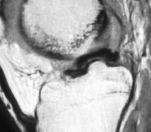
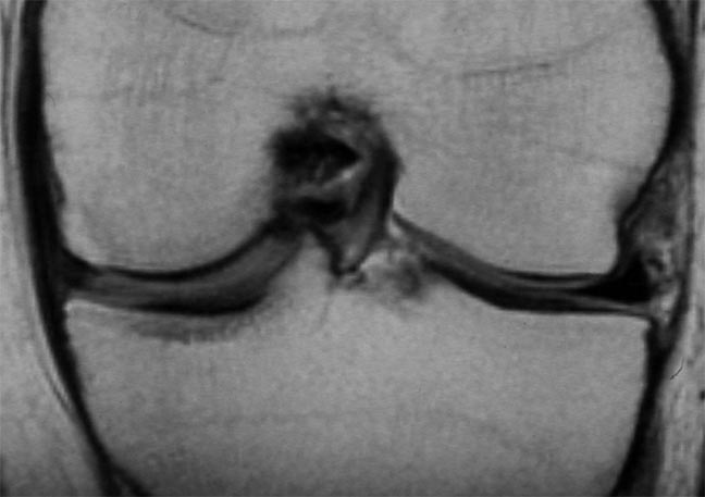
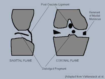
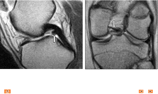
- Pathology: Meniscal bucket handle
tear, with inner meniscal fragment displaced into the intercondylar
notch, anterior to the PCL.
Double PCL sign occurs with medial meniscal tears only; the ACL
prevents displacement of lateral meniscal fragments into the
intercondylar notch, anterior to the PCL.
- Imaging findings: Band of low
signal intensity, anterior and parallel to the PCL, simulating the PCL
(sagittal view). Band of low
signal intensity within the intercondylar notch, between the PCL and
tibial plateau; associated truncation of triangular configuration of
peripheral meniscus (coronal view).
- Note: The double PCL sign tear may be considered as a subset of
fragment-in-notch type medial meniscal tears, in which the displaced
fragment lies in the same sagittal plane as the PCL.
|
|
17
|
|
|
18
|
|
|
19
|
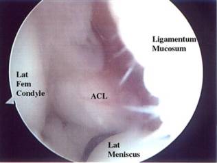
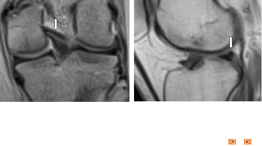
- Pathology: Meniscal bucket handle
tear, with inner meniscal fragment displaced into the intercondylar
notch, anterior and lateral to the ACL.
Double ACL sign commonly occurs with lateral meniscal bucket
handle tears.
- Imaging findings: Band of low
signal intensity, anterior and lateral to the ACL (sagittal view). Because of the oblique orientation of
the ACL, the meniscal fragment may parallel and simulate the ACL when
viewed on oblique sagittal orientation.
Band of low signal intensity may be seen within the intercondylar
notch, parallel to the ACL; associated truncation of triangular
configuration of peripheral meniscus (coronal view).
- Note: The double ACL sign tear may be considered as a subset of
fragment-in-notch type meniscal tears, in which the displaced fragment
lies in the same sagittal plane as the PCL.
|
|
20
|
|
|
21
|
|
|
22
|
|
|
23
|
- The following variants may mimic bucket-handle tears of the meniscus:
- TRANSVERSE LIGAMENT
- MENISCOFEMORAL LIGAMENT (Humphry)
- MENISCOFEMORAL LIGAMENT (Wrisberg)
- POPLITEUS TENDON
- INTRAARTICULAR LOOSE BODY
- LATERAL INFERIOR GENICULATE ARTERY—mimics anterior horn tear
|
|
24
|
|
|
25
|
|
|
26
|
|
|
27
|
|
|
28
|
|
|
29
|
- Gentili A, Seeger L, et al.
Anterior cruciate ligament tear: indirect signs at MR imaging. Radiology
1994;193:835-840.
- Helms CA, Laorr A, Cannon WD. The
absent bow tie sign in bucket-handle tears of the menisci in the
knee. AJR 1998;170(1):57-61.
- Mesgarzadeh M, Moyer R, et al. MR
imaging of the knee: expanded classification and pitfalls to
interpretation of meniscal tears.
RadioGraphics 1993;13:489-500.
- Murata Y, Yoshida D, et al. MRI of meniscal bucket handle tear: the
double PCL versus the double ACL.
Abstract ECR 2000 meeting.
- Ruff C, Weingardt JP, et al. MR
imaging patterns of displaced meniscus injuries of the knee. AJR 1998;170(1):63-7.
- Singson RD, Feldman F, et al. MR
imaging of displaced bucket-handle tear of the medial meniscus. AJR
1991;156(1):121-4.
- Sparacia G, LoCasto A, et al.
Bucket-handle tears of the knee menisci: pitfall in
interpretation at MR imaging.
RSNA presentation/paper 1997.
- Weiss K, Morehouse H, Levy M.
Sagittal MR images of the knee; a low signal band parallel to the
posterior cruciate ligament caused by a displaced bucket handle tear. AJR
1991;156:117-199.
- Wright DH, De Smet AA, Norris M.
Bucket-handle tears of the medial and lateral menisci of the
knee: value of MR Imaging in detecting displaced fragments. AJR
1995;165:621-5.
- Vahlensieck M, Genant H, Reiser M.
MRI of the musculoskeletal system. Injury 2002;33(2):191.
|
 Notes
Notes