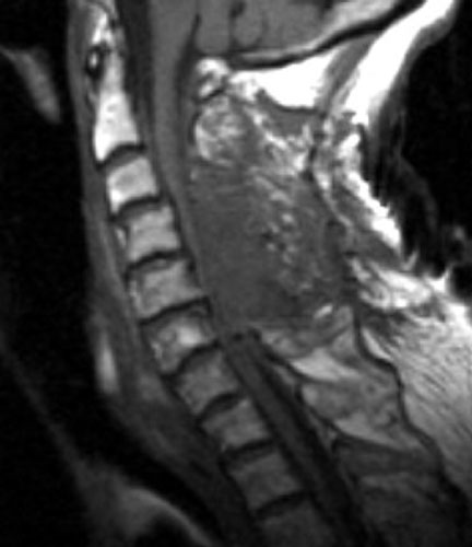
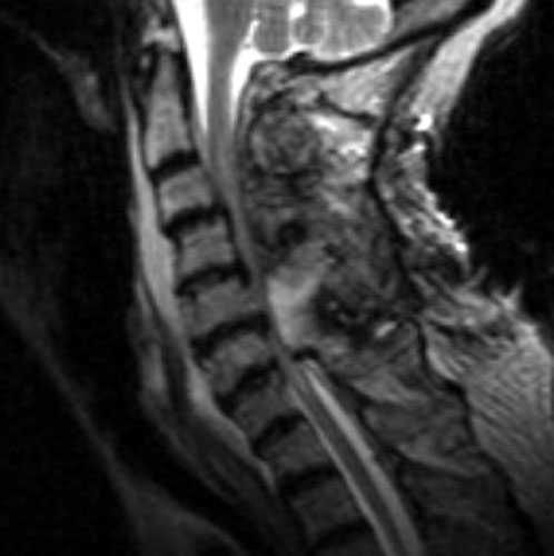
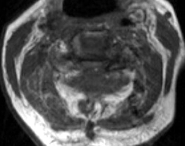
| Non-enhanced sagittal T1 (A), sagittal T2 (B) and axial T1
images demonstrate a large fluid collection,
predominantly low signal on T1, bright on T2 with a few areas of T1 hyperintesity
mixed within. These constellation of findings are consistent with a
post-operative epidural hematoma with blood products of various ages. There is
severe severe spinal canal stenosis at C3-4, and very severe spinal canal
stenosis at C4-5 and C5-6. The cord is markedly flattened in the
anterior-posterior dimension, to approximately 2-3 mm secondary to mass effect
from this fluid collection. There is abnormal T2 weighted hyperintensity within
the substance of the cord at the C5-6 and C6-7 levels, consistent with some
edema. |
 A
A
 B
B
 C
C