|
Up
Wound Infection
Seroma and Epidural Scar
Epidural Hematoma - CT
Epidural Hematoma - MRI
Instability
Graft Reabsorption
Endplate Fracture
Graft Collapse
Nonunion
Displaced Strut Graft
Broken Screw
Screw in Spinal Canal
Slipped rod
Screw in Disc space
Screw Loosening & Hematoma
Screw Backing Out
Screw Loosening
| |
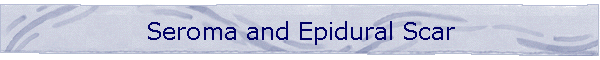
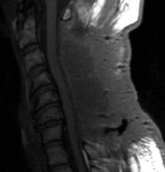
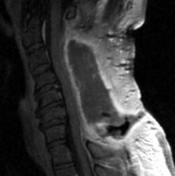
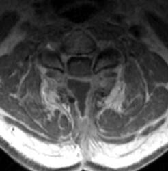
Seroma and Epidural Scar
46 y.o. male, initial complaint of progressed with myelopathy,
difficulty walking, and imbalance. Underwent laminectomy C3 to C7, 2 months
prior to these images. Worsening numbness and weakness brought patient to seek
neurosurgical consultation again, prompting repeat imaging.
 A A |
 B B |
 C C |
 D D |
|
Click on the images to view a larger version!
|
| Sagittal T1 pre (A) and post contrast
(B), sagittal T2 (C) and Axial T1 post contrast (D) MRI images
of the cervical spine demonstrating a large posterior fluid collection with an
obvious sinus tract inferiorly and associated gas bubbles. This was proved to be
a post-operative seroma and epidural scar (note the thick rind of enhancement)
that was partially accounting for this patient's residual and worsening symptoms
despite the initial decompressive surgery. |
|
|
|