Compression fractures of the spine are common in elderly and osteoporotic patients. They result from anterior or lateral flexion. The typical appearance is loss of height of the anterior aspect of the vertebral body with preservation of the posterior elements and generally the posterior aspect of the vertebral body. On the frontal view, there may be subtle deviation of the paraspinous line due to edema. Differentiation from a pathologic fracture of the spine due to a metastasis is usually of clinical concern, but simple compression fractures are usually due to osteoporosis.
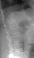 A A | Lateral radiograph of the lumbar spine. This shows an osteoporotic compression fracture of the L1 vertebral body. |
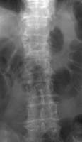 B B | AP radiograph of the lumbar spine. This shows loss of height of the L1 vertebral body, corresponding to a compression fracture. |
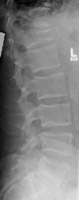 C C | Lateral radiograph of the lumbar spine. This is an image from a different patient and shows a severe compression fracture of the T12 vertebral body. |
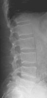 D D | Lateral radiograph of the lumbar spine. Two years prior there was osteopenia, but the fracture was not present. |
 A
A B
B C
C D
D